
Dental caries, colloquially known as cavities, are a pervasive and insidious threat to oral health. Understanding what a cavity looks like in its nascent stages, and how it progresses, is paramount for proactive intervention and preservation of your pearly whites. A cavity is not merely a superficial blemish; it’s a microscopic battleground where bacterial armies wage war against the enamel fortress protecting your teeth.
The Visual Manifestations: A Stage-by-Stage Unveiling
Initially, cavities are almost phantom-like, existing as subsurface demineralization imperceptible to the naked eye. Think of it as an iceberg, where the most significant damage lurks beneath the surface, unseen and potentially devastating.
1. The Enamel’s Whisper: White Spots
The very first sign is often chalky white spots on the enamel surface. These spots represent areas where the enamel has begun to lose minerals due to acid erosion from bacterial plaque. These lesions are not always indicative of active decay. Sometimes, these spots, also termed “incipient lesions”, can be remineralized with diligent oral hygiene and fluoride application. Detecting these requires meticulous examination and good lighting. It’s like spotting the faintest constellation in a night sky; you need to know where to look and have the right conditions.
2. The Subsurface Intrusion: A Subtle Shading
As the demineralization progresses, the white spots may darken slightly, becoming a dull, opaque hue. This indicates that the acid has penetrated deeper into the enamel’s crystalline structure, creating microporosities. The tooth’s structural integrity is being compromised. This early stage is crucial, offering a window of opportunity for minimally invasive treatments like fluoride varnishes or resin infiltration.
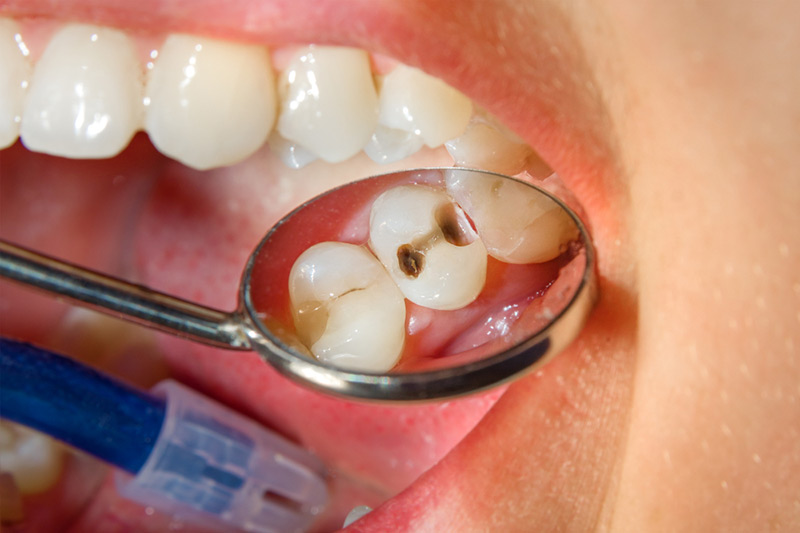
3. The Breakthrough: A Visible Pit or Hole
Once the enamel’s outer layer is breached, a visible pit or hole forms. This is the classic image of a cavity that most people recognize. The size and shape of the cavity will vary depending on the location and the rate of progression. These imperfections are often jagged and irregular, unlike the smooth contours of healthy enamel.
4. The Dentin’s Distress: A Change in Color and Texture
Beneath the enamel lies the dentin, a softer, more porous tissue. Once the cavity reaches the dentin, it tends to spread more rapidly. The affected dentin typically appears darker than healthy dentin, ranging from yellow-brown to almost black. The texture also changes, becoming soft and leathery to the touch, easily excavated with a dental instrument.
5. The Pulpal Peril: Pain and Inflammation
If left untreated, the cavity can eventually reach the dental pulp, the innermost layer of the tooth containing nerves and blood vessels. This can lead to significant pain, sensitivity to temperature changes, and inflammation of the pulp (pulpitis). In severe cases, the infection can spread beyond the tooth, resulting in an abscess.
Beyond the Visual: Ancillary Indicators
While visual inspection is crucial, there are other indicators that might suggest the presence of a cavity.
1. Sensitivity to Temperature and Sweetness: A sharp, fleeting pain when consuming hot, cold, or sweet foods is a common symptom. This sensitivity arises from the exposure of dentinal tubules, which transmit stimuli directly to the pulp.
2. Pain Upon Biting or Chewing: As the cavity grows larger, it can weaken the tooth’s structure, making it painful to bite down or chew on that side of the mouth.
3. Food Impaction: A cavity can create a niche where food particles become trapped, leading to localized inflammation and discomfort.
4. Halitosis (Bad Breath): The bacteria thriving within a cavity can produce volatile sulfur compounds, contributing to persistent bad breath.
5. A Rough or Sharp Edge: Running your tongue along the tooth may reveal a rough or sharp edge where the enamel has been eroded.
Differential Diagnosis: Distinguishing Cavities from Mimickers
It’s important to differentiate cavities from other conditions that can mimic their appearance. Staining from coffee, tea, or tobacco can create discoloration that resembles early decay. Dental fluorosis, a condition caused by excessive fluoride intake during tooth development, can result in white spots on the enamel. However, unlike cavities, these conditions do not typically involve structural damage to the tooth.
The Imperative of Early Detection: A Call to Action
The earlier a cavity is detected, the less invasive the treatment required. Early intervention, such as fluoride therapy or resin infiltration, can often halt the progression of the decay and prevent the need for a filling. Regular dental check-ups, including professional cleanings and radiographic examinations, are essential for identifying cavities in their nascent stages. Think of your dentist as a cartographer, mapping the terrain of your mouth to identify and address any emerging threats before they escalate.
In conclusion, understanding the multifaceted appearance of cavities, from the subtle whisper of white spots to the blatant declaration of a visible pit, is crucial for maintaining optimal oral health. Proactive monitoring, coupled with meticulous oral hygiene and regular dental visits, empowers you to defend your teeth against the insidious onslaught of dental caries. Remember, the battle for a healthy smile is won not in the dental chair, but in the daily rituals of prevention and care.


Leave a Comment