
Plantar warts, verrucae plantares in medical parlance, are a common affliction, particularly among those who frequent communal bathing facilities or engage in activities that involve barefoot ambulation. Understanding their morphology and distinguishing them from other cutaneous lesions is paramount for appropriate management. This exposition delves into the multifaceted characteristics of plantar warts, elucidating their visual attributes, tactile qualities, and potential complications.
I. Macroscopic Appearance: The Deceptive Simplicity
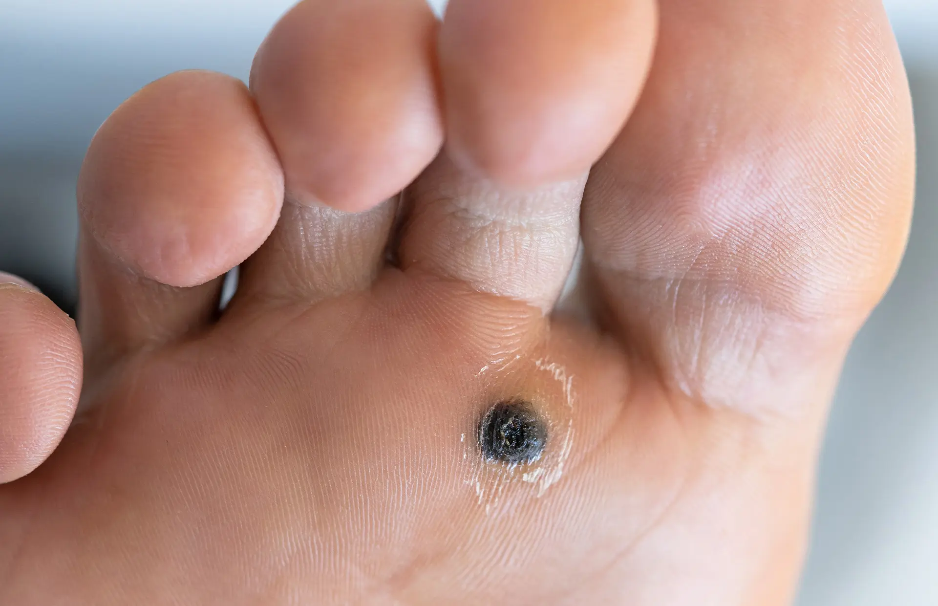
At first blush, a plantar wart may appear unremarkable, often mistaken for a callus or corn. A closer inspection, however, reveals key differentiators. The cardinal sign is the presence of tiny black dots within the lesion. These punctate hemorrhages, often described as “wart seeds,” are thrombosed capillaries, the remnants of blood vessels that supplied the wart tissue. The surface of the wart is typically rough and uneven, exhibiting a cauliflower-like texture. Unlike calluses, which tend to follow the natural lines of the skin, plantar warts disrupt these lines, creating a noticeable discontinuity.
II. Palpatory Examination: Feeling for Subtleties
Tactile assessment provides additional clues. Plantar warts are typically tender to direct pressure. Lateral compression, squeezing the wart from the sides, often elicits a more pronounced pain response than applying pressure from above. This is because the lateral pressure stimulates the nerve endings surrounding the lesion. Calluses, conversely, are generally less sensitive to lateral compression.
III. Variants and Subtypes: A Spectrum of Presentations
The presentation of plantar warts can vary depending on factors such as the individual’s immune response, the duration of the infection, and the location on the foot. Several distinct subtypes exist:
A. Solitary Wart: A single, well-defined lesion. Often the initial presentation, it can serve as a seeding point for the development of satellite warts.
B. Mosaic Wart: A cluster of closely packed, small warts that coalesce to form a larger, plaque-like area. These are notoriously resistant to treatment due to their extensive nature.
C. Subungual Wart: A wart that develops under the toenail. These can be particularly painful and difficult to treat due to their location and the potential for nail plate involvement.
D. Periungual Wart: A wart that develops around the toenail. Similar to subungual warts, they can be challenging to manage.
IV. Differential Diagnosis: Distinguishing Warts from Imitators
Several other conditions can mimic the appearance of plantar warts, necessitating a thorough clinical evaluation. Important considerations include:
A. Calluses and Corns: As mentioned earlier, these are areas of thickened skin caused by repeated pressure or friction. Unlike warts, they typically follow skin lines and lack the characteristic black dots.
B. Foreign Body Granulomas: These are inflammatory reactions to foreign materials embedded in the skin. They can sometimes resemble warts, but a history of trauma or penetration may be present.
C. Squamous Cell Carcinoma: In rare cases, a persistent or atypical plantar lesion may represent a cutaneous malignancy. A biopsy is often necessary to rule out this possibility, especially in individuals with a history of skin cancer or sun exposure.
D. Plantar Fibromatosis (Ledderhose Disease): This condition involves the development of fibrous nodules in the plantar fascia. These nodules are typically firm and deep, lacking the superficial characteristics of warts.
V. Diagnostic Adjuncts: When Visual Inspection Isn’t Enough
In some cases, the diagnosis may not be clear based solely on clinical examination. Adjunctive diagnostic tools can provide further clarification:
A. Dermoscopy: A non-invasive technique that uses a magnifying lens and polarized light to visualize subsurface skin structures. Dermoscopy can enhance the visualization of thrombosed capillaries and other characteristic features of warts.
B. Shave Biopsy: A superficial scraping of the lesion can be examined under a microscope to confirm the presence of koilocytes, characteristic cells infected with human papillomavirus (HPV).
C. Polymerase Chain Reaction (PCR): A molecular test that can detect the presence of HPV DNA in a tissue sample. This can be useful for identifying specific HPV types and confirming the diagnosis.
VI. Potential Complications: Beyond the Pain
While plantar warts are generally benign, they can lead to several complications if left untreated:
A. Pain and Discomfort: Plantar warts can cause significant pain, particularly with weight-bearing activities. This can lead to altered gait patterns and secondary musculoskeletal problems.
B. Secondary Infection: The disrupted skin surface can provide a portal of entry for bacteria, leading to secondary infections such as cellulitis.
C. Spread: Warts are contagious and can spread to other areas of the body or to other individuals through direct contact.
D. Psychological Impact: The unsightly appearance of warts can be distressing and lead to social anxiety, particularly in children and adolescents.
VII. Conclusion: A Multifaceted Understanding
The appearance of a plantar wart is more than meets the eye. It requires a discerning assessment of its macroscopic features, tactile qualities, and potential variations. Differentiating it from other conditions, such as calluses, foreign body granulomas, and even rare malignancies, is crucial. Understanding the potential complications associated with plantar warts emphasizes the importance of prompt and appropriate management. By possessing a comprehensive understanding of the visual and tactile characteristics of these lesions, clinicians and individuals alike can better navigate the diagnostic and therapeutic landscape of plantar warts.


Leave a Comment